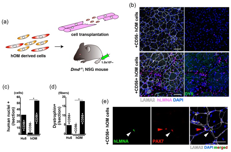
Figure 5.
CD56-positive cells from orbicularis oculi muscle have regenerative capacity for Duchenne muscular dystrophy model mice. (a) A schematic model for the transplantation with sorted cells of orbicularis oculi muscle (hOM) into DMD-null (Dmd-/y) and NSG mice. (b) Immunostaining for LAMININ alpha-2 (LAMA2, labelled with Alexa647, white), human nuclear LAMIN A/C (hLMNA, labelled with Alexa594, pink), and DYSTROPHIN (DYS, labelled with Alexa488, green) in tibialis anterior (TA) muscles of Dmd-/y. NSG mice were injected with CD56-negative (upper panels) or CD56-positive (lower panels) cells derived from the orbicularis oculi 2 weeks after intramuscular engraftment. Total nuclei were stained with DAPI (blue). Scale bar, 50 μm. (c) The quantification of transplanted human nuclear cells (human nuclei+) in TA muscles engrafted with equal numbers of immortalized human myoblast Hu5 cells, and CD56-negative (+CD56−) or positive (+CD56+) cells from orbicularis oculi. n = 3 independent replicates; p-values are determined by t-test from a two-tailed distribution. * p < 0.01. Error bar indicates ±SEM. (d) The quantification of total DYSTROPHIN-positive regenerative myofibers on the section transplanted with equal numbers of indicated cells as in (c). n = 3 independent replicates; p-values are determined by t-test from a two-tailed distribution. * p < 0.01. Error bar indicates ±SEM. (e) Immunostaining for PAX7 (labelled with Alexa594, red), hLMNA (labelled with Alexa488, green), LAMA2 (labelled with Alexa647, white), and DAPI (blue) on a section of engrafted tibialis anterior with cultured CD56-positive cells from the orbicularis oculi. White arrow shows a transplanted PAX7+ cell. Red arrow shows intact mouse muscle satellite cell. Scale bar, 50 μm.

