Fig. 1.
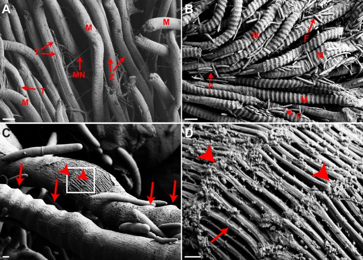
Invasion of mandibular muscle space by Ophiocordycepskimflemingiae – evidence for muscle over-contraction. Infected mandibular muscle displays unique characteristics as visualized by scanning electron microscopy (SEM) (controls, n=3; infected, n=6). (A) In uninfected controls, mandibular muscle cells demonstrate an even morphology with regularly distributed striations (z-lines). Motor neurons and tracheoles are abundant. Scale bar: 20 µm. M, muscle; MN, motor neuron; T, tracheole; Z, z-line. (B) Fungal cells completely invade the inter-muscle space and are in close contact with individual muscle cells. Infected muscle cells demonstrate a unique morphology wherein the z-lines appear to be swollen and the sarcomeres shortened, giving the regular striations a very pronounced appearance. Scale bar: 20 µm. M, muscle; F, fungus. (C) As a result of extensive damage to the sarcolemma, individual myofibrils underneath the membrane are exposed in infected muscle. Areas of sarcomere shortening (arrows: swollen, unexposed regions; arrowheads: exposed regions) are evident. Scale bar: 20 µm. (D) Magnification of the boxed region in C. Presumed z-lines are indicated by arrowheads while individual myofibrils are indicated by arrows. The z-lines display a frayed, damaged appearance. Scale bar: 1 µm.

