Fig. 3.
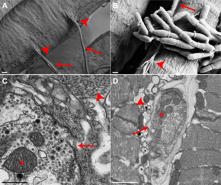
Motor neurons and neuromuscular junctions are maintained in infected mandibular muscle at the time of biting. In control ants (A), numerous motor neurons (arrows) and neuromuscular junctions (NMJs, arrowheads) are evident along the length of individual muscle cells (SEM image). Scale bar: 2 µm. (B) Motor neurons (arrow) and NMJs (arrowhead) in infected mandibular muscle are maintained (SEM image). In some instances, these structures are in close contact with fungal cell bodies. Scale bar: 2 µm. In both control (C) and infected (D) ants, nervous tissue is present near and/or in contact with muscle, as identified by TEM (nervous tissue, arrows; muscle cells, arrowheads). M, mitochondria. Scale bars: C, 500 nm; D, 1 µm. Controls, n=3; infected, n=6.

