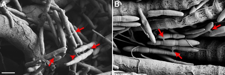
Fig. 5.
Ophiocordycepskimflemingiae fungal cells demonstrate collective behavior via anastomosis tube formation. (A,B) SEM reveals that, within infected muscle, individual O. kimflemingiae cells form extensive networks with each other, connected via the formation of anastomosis tubes (arrows). In many instances, numerous anastomosis tubes form along the length of one fungal cell, and formation of new tubes (A, arrowhead) is evident. Scale bars: 10 µm. Controls, n=3; infected, n=6.

