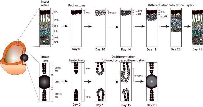
Fig. 4.
Regeneration of ocular tissues. (Top) Following retinectomy (detachment of the RPE from the photoreceptor cell layer), a new pigmented cell layer appears first. This is followed by the formation of neuro-retinal cell types in an order that recapitulates development: ganglion cells form first, followed by amacrine cells, horizontal cells and Müller glia. (Bottom) The lens can regenerate following lentectomy (lens removal), via the dedifferentiation and subsequent transdifferentiation of pigmented epithelial cells of the dorsal iris. Newts retain lens regeneration ability throughout adulthood, unlike axolotls, in which the ability to regenerate the lens is lost 2 weeks after hatching. dIPE, dorsal iris pigmented epithelium; dIPESCs, dorsal iris pigmented epithelium cells; GCL, ganglion cell layer; INL, inner nuclear layer; ONL, outer nuclear layer; OPL, outer plexiform layer; proNR, inner rudimentary layer; proRE, retinal pigmented epithelium rudimentary layer; RPE, retinal pigmented epithelium; RPESCs, retinal pigmented epithelium stem cells; vIPE, ventral iris pigmented epithelium.

