
Fig. 5.
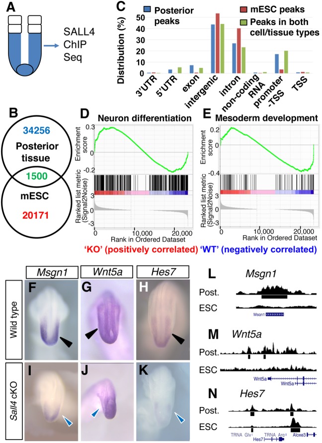
SALL4 ChIP-Seq analysis of the posterior tissue and downregulation of mesoderm differentiation genes in Sall4 cKO embryos. (A) Schematic of dissected posterior tissue (blue) for SALL4 ChIP-Seq analysis. (B) Venn diagram for SALL4-enriched sequence numbers in the posterior tissue and mESCs. (C) Distributions of SALL4-enriched sequences in the posterior tissue, mESCs and both cell/tissue types. (D,E) GSEA analysis of genes with GO terms of neural differentiation (D) and mesoderm development (E) among genes that are enriched by SALL4 in the posterior tissue. (F-K) Whole-mount in situ hybridization of the indicated mesodermal genes in WT and Sall4 cKO embryos at E9.5. Black arrowheads point to normal expression in WT PSM (F-H). Blue arrowheads point to reduced expression in the PSM in Sall4 cKO embryos (I-K). (L-N) Visual representation of SALL4 ChIP-Seq results of the indicated gene loci. Post., the posterior tissue.