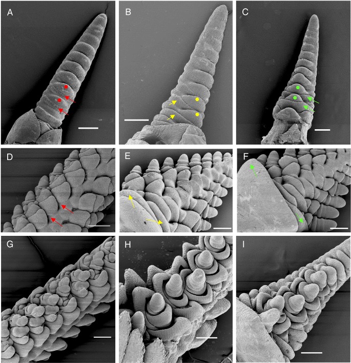
Fig. 3.
Scanning electron microscopy images. Early double-ridge stage (A-C) and later stage (D-I) showing the fate of the lateral meristems. (A) Kronos control. (D,G) vrn1-null control. Red arrows indicate the repressed lower leaf ridge, and red dots the upper ridges that develop into normal spikelets (D,G). (B,E,H) vrn1ful2-null mutants. Yellow arrows indicate the partially repressed lower leaf ridges that develop into bracts (see Fig. 2D) and yellow dots indicate the upper ridges that develop into intermediate meristems that generate tiller-like structures with altered floral organs (see Fig. 2E-G). (C,F,I) vrn1ful2ful3-null mutants. Green arrows indicate basal lower leaf ridges that develop into normal leaves (see Fig. 2H) and green dots indicate upper ridges that produce lateral vegetative meristems that generate vegetative tillers with no floral organs (see Fig. 2I-J). Scale bars: 200 µm.

