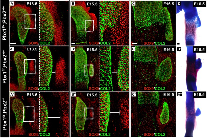
Fig. 7.
Pbx1 and Pbx2 coordinately regulate the patterning of proximal superstructures. (A-C″) Sagittal sections through the proximal humeri of E13.5 (A-A″), E15.5 (B-B″) and E16.5 (C-C″) limbs from control (A-C), Prx1-Cre;Pbx1floxed (B-B″) or Prx1-Cre;Pbx1floxed;Pbx2het (C-C″) embryos that were stained against SOX9 and COL2A1. In A-B″, the right side of the panel is an enlargement of the humerus-deltoid tuberosity boundary region demarcated by a white rectangle on the left. At E13.5, spatial organization of deltoid tuberosity precursors in both mutants was abnormal (A′,A″) as deltoid tuberosity precursors were scattered laterally away from the humeral shaft. The gap between the humeral shaft and deltoid tuberosity precursors was twice as large in Prx1-Cre;Pbx1floxed;Pbx2het mutants than in Prx1-Cre;Pbx1floxed mutants (A′,A″, white brackets). At E15.5, whereas cells at the humerus- deltoid tuberosity boundary of Prx1-Cre;Pbx1floxed mutants began to express high levels of SOX9, such expression was not observed in Prx1-Cre;Pbx1floxed;Pbx2het mutants. Moreover, the difference in boundary region size remained consistent (B,B′, white brackets). At E16.5, deltoid tuberosity morphology ranged from attached but aplastic deltoid tuberosity in Prx1-Cre;Pbx1floxed mutants (C′) to a detached deltoid tuberosity in Prx1-Cre;Pbx1floxed;Pbx2het mutants (C″). (D-D″) Skeletal preparations of E16.5 forelimbs further validate these diverging morphologies. Scale bars: 100 µm.

