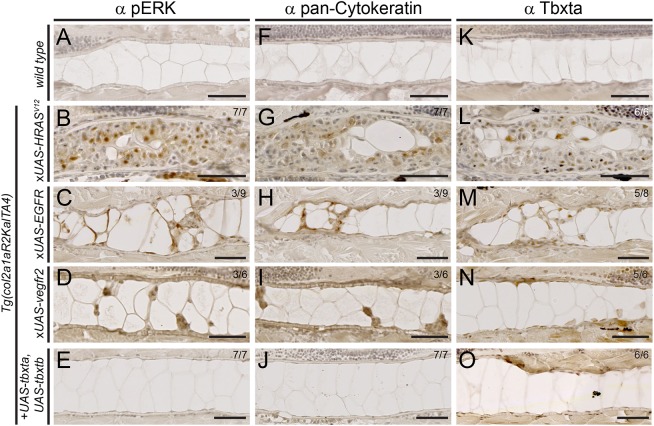
Fig. 4.
Expression of chordoma markers in RTK-transformed zebrafish notochords. (A-O) Immunohistochemistry on sagittal sections through the notochord of 5 dpf zebrafish embryos of the indicated genotypes, expressing either stable or mosaic transgenes. (A-E) MAPK pathway activation through HRASV12, EGFR and kdr overexpression results in nuclear pERK staining in the notochord, whereas controls and tbxta,tbxtb-injected embryos are negative for pERK staining. (F-J) HRASV12, EGFR and kdr overexpression results in staining for pan-Cytokeratin, whereas tbxta,tbxtb-injected embryos are negative for pan-Cytokeratin staining akin to wild-type controls. (K-O) Whereas control notochords show faint to no Tbxta signal, owing to low cell density of the sheath layer (K), HRASV12-overexpressing notochords (L) as well as EGFR-overexpressing (M) and kdr-overexpressing (N) notochords show prominent nuclear Tbxta staining. The tbxta,tbxtb-overexpressing notochords also stain positive (O), confirming transgenic tbxta expression. Scale bars: 50 µm. See also Fig. S4.

