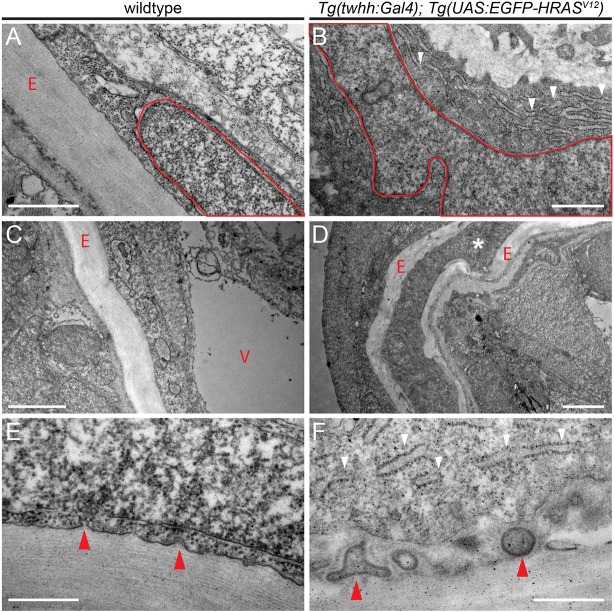
Fig. 6.
Aberrant ECM and ER accumulation in HRASV12-induced zebrafish chordomas. (A-F) Transverse sections through wild-type (A,C,E) and HRASV12-transformed (B,D,F) zebrafish notochord at 8 dpf imaged using TEM. (A,B) Wild-type cells have a pill-shaped, regular nucleus (red outline, A) and form regular ECM layers (red letter ‘E’) secreted by the sheath cells at the outside of the notochord. Nuclei in transformed cells expand and develop lobed and distorted nuclear shapes (red outline, B); transformed cells further accumulate extensive ER lumen (white arrowheads, B). (C,D) In wild-type notochords, vacuolated cells (red letter ‘V’) take up the majority of the space inside the ECM-lined notochord (red letter ‘E’) to provide mechanical stability; transformed notochords become filled with non-vacuolated cells and secrete aberrant amounts of ECM that leads in extreme cases to entombed cells trapped in ECM layers (white asterisk, D). (E,F) Membrane details of wild-type versus transformed notochord sheath cells. Wild-type notochords show budding of vesicles that transport collagen for stereotypically layered ECM build up (red arrowheads); in transformed notochords, the secretion process appears to be overactive (ER accumulation shown by white arrowheads, F) and results in membrane inclusions within the ECM (red arrowheads). Scale bars: 1 μm (A,B); 2 μm (C,D); 0.5 μm (E,F). See also Fig. S6.

