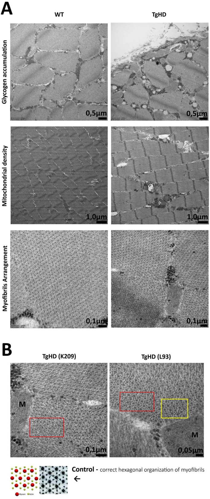
Fig. 1.

Ultrastructure of skeletal muscle from 48-month-old WT and TgHD minipigs analyzed using transmission electron microscopy. (A) Accumulation of glycogen, mitochondrial density and myofibril arrangement in WT and TgHD muscle. Glycogen is indicated in TgHD muscle with an asterisk (top). (B) Higher mitochondrial density and local disruption of hexagonal organization of myofibrils is observed in TgHD animals (red boxes). The amount of actin is also increased (yellow box); M, mitochondrion. The selected pair represents the results of the entire group. TgHD, transgenic; WT, wild type; K209, L93, animal identification numbers.
