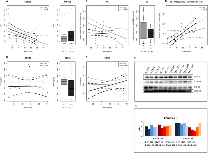
Fig. 4.
The time course impairment of function and protein expression for complex II in TgHD minipig skeletal muscle. (A) Ratio of complex II to citrate synthase activity (SQR/CS parameter) is significantly increased in TgHD animals compared with WT animals (P=0.125). (B) Complex II-dependent respiration is significantly decreased in TgHD animals (P=0.0008). (C) Rate of oxidation of [1,4-14C]succinate in TgHD animals shows an increasing trend and a different slope in comparison to WT animals, which may indicate the need for increased turnover for RCCII. (D) Expression of subunit SDH30 of RCCII is significantly lower in TgHD animals than in WT animals (P=0.0132). Reduced protein content was observed for all age categories. (E) In contrast, the SDH70 subunit of RCCII shows a rising trend, but the groups did not differ significantly. Intensity in panels D and E represents the signal of proteins analyzed by western blot and quantified using the Quantity One 1-D Analysis Software (Bio-Rad). In the box-whisker plots, median and quartiles are displayed. Circles represent outliers that are further than 1.5IQR from the corresponding quartile. Whiskers show the range of non-outliers. Boxes represent the TgHD and WT groups that consist of all animals in all ages between 24 and 66 months; numbers of analyzed samples are indicated. (F) Representative WB analysis of subunits SDH30 and SDH70 at the age of 66 months. The respective protein pairs from the same blot are indicated with a bracket. Analyses were conducted for samples collected at the ages of 24, 36, 48 and from 66 months. (G) Quantification of the normalized western blot signal from 5 TgHD and 5 WT muscles (shown in panel F) using Quantity One 1-D Analysis Software (Bio-Rad). Enzyme activities were measured spectrophotometrically, respiration was measured by high-resolution respirometry on an OROBOROS oxygraph and the protein content was analyzed using specific antibodies (Abcam) for western blotting.

