Fig. 6.
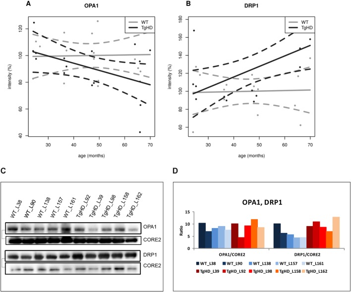
Differential expression of selected mitochondrial membrane proteins in TgHD minipig skeletal muscle during aging. (A) OPA1 expression showed a decreasing trend with age in TgHD animals compared with WT animals. (B) DRP1 expression showed an increasing trend in TgHD animals compared with WT animals. Western blot analyses were conducted for samples collected at the ages of 24, 36, 48 and 66 months. Intensity in panels A and B represents the signal of proteins analyzed by western blot and quantified using the Quantity One 1-D Analysis Software (Bio-Rad). (C) Representative western blot analyses of OPA1, DRP1 and CORE2 at the age of 66 months; the respective protein pairs from the same blot are indicated with a bracket. (D) Quantification of the normalized western blot signal from 5 TgHD and 5 WT muscles (shown in panel C) using Quantity One 1-D Analysis Software (Bio-Rad).

