Fig. 1.
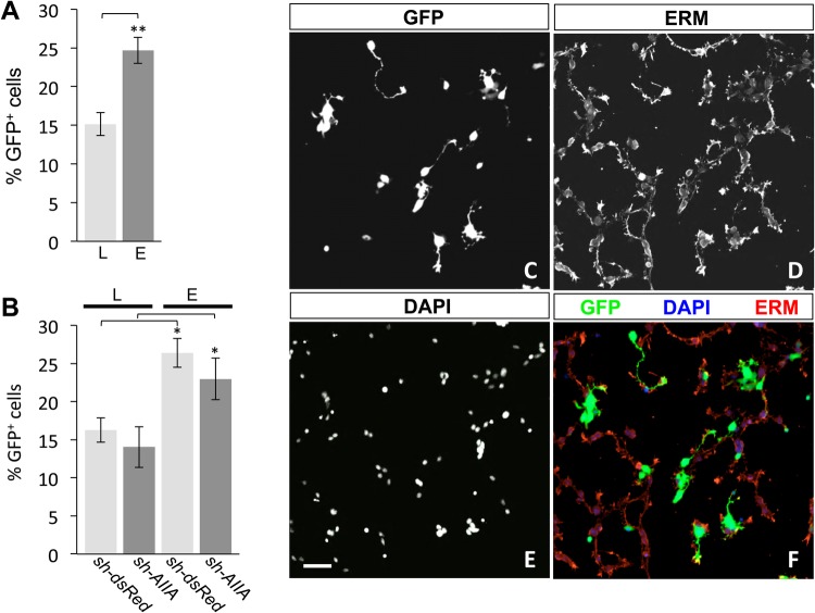
Transfection of shRNA vectors in DSC neurons by lipofection and electroporation. (A) Combined transfection efficiency assessed by the percentage of GFP+ neurons (mean±s.e.m.) in dsRedΔDSC and AIIAΔDSC cultures by lipofection (L, 15.2%±1.5%) or electroporation (E, 24.7%±1.7%). Electroporation significantly improved transfection efficiency compared with lipofection (**P<0.002, n=3) (Student's two-tailed t-test). (B) Transfection efficiencies (% GFP+ neurons; mean±s.e.m.; n=3 for each condition) for dsRedΔDSC and AIIAΔDSC cultures show no vector-specific differences in neurons transfected by either lipofection (dsRedΔDSC, 16.3%±1.6%; AIIAΔDSC, 14.0%±2.7%) or electroporation (dsRedΔDSC, 26.4%±1.9%; AIIAΔDSC, 23.0%±2.7%). In contrast, transfection efficiencies revealed that in dsRedΔDSC and AIIAΔDSC cultures, the percentage of GFP+ neurons increased by 62% and 63%, respectively, in electroporated cultures compared with transfection by lipofection (*P<0.05, n=3 for both conditions) (Student's two-tailed t-test). (C–F) Dissociated dsRedΔDSC culture labeled for (C) GFP, (D) ERM and (E) DAPI. Merged image (F) shows GFP expression in electroporated neurons (green), ERM labeling localized to the cellular membranes (red) and DAPI staining the nuclei (blue). Scale bar: 50 μm.

