Fig. 2.
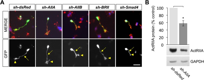
Growth cone expression of type II BMP receptor-targeted shRNA vectors in DSC neurons. (A) Representative images of DSC neurons electroporated with the indicated shRNA vectors in dissociated culture labeled for GFP (green), ERM (red) and DAPI (blue). The presence of GFP reflects expression of each of the shRNA vectors in cultured DSC neurons including in the axonal processes (arrows) and growth cones (arrowheads). In the far right column, a Smad4ΔDSC neuron is shown, treated with 50 ng/ml BMP7 for 30 min, and represents a collapsed growth cone (arrowhead). Scale bar: 50 μm. (B) Western blots of dsRedΔDSC or AIIAΔDSC lysates probed with an anti-ActRIIA antibody. Detection of GAPDH provided a loading control. Electroporation of sh-AIIA in DSC cultures decreases ActRIIA protein expression to 58%±7.4% of the expression levels in GFP-enriched, dsRedΔDSC lysates (mean±s.e.m.; n=4; *P<0.05) (Student's two-tailed t-test).

