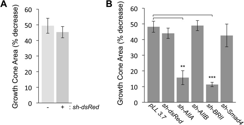
Fig. 3.
BMP7-evoked growth cone collapse in DSC neurons in the presence of control and type II BMP receptor shRNA vectors. (A) The percentage of growth cone area decrease [GCAD=(100–{[(GC area in the presence of BMP7–control GC area)/control GC area]×100}); mean±s.e.m.] in dsRedΔDSC neurons stimulated by 50 ng/ml BMP7 for 30 min is not affected by expression of the non-specific shRNA vector, sh-dsRed (GCAD=48.3%±3.3%) when compared with untransfected, GFP− neurons (GCAD=49.4%±4.8%) in the same culture (n=3). (B) Growth cone area decrease (mean±s.e.m.) in response to 50 ng/ml BMP7 was measured for shRNA-transfected GFP+ neurons only. Expression of pLL3.7, the shRNA parent vector, (GCAD=48.2%±3.5%; n=3) or knockdown of ActRIIB (AIIBΔDSC, GCAD=54.4%±3.3%; n=3) or Smad4 (Smad4ΔDSC, GCAD=42.6%±7.2%; n=3) did not interfere with BMP7-evoked growth cone collapse. In contrast, BMP7-evoked growth cone collapse was significantly inhibited in AIIAΔDSC (GCAD=15.7%±4.4%; n=4, **P<0.005) and BRIIΔDSC cultures (GCAD=11.3%±1.4%; n=3, ***P<0.001) (Student's two-tailed t-test).

