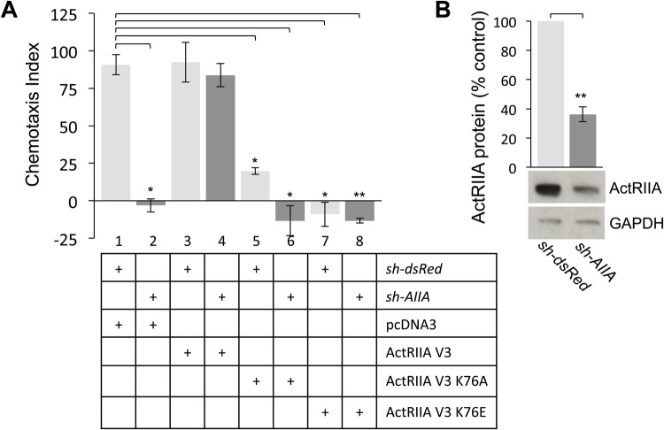
Fig. 5.
ActRIIA V3 K76 receptor variants cannot rescue sh-AIIA-mediated inhibition of BMP7-stimulated chemotaxis. (A) Chemotaxis in response to 50 ng/mL BMP7 (CI={[(no. BMP7 treated cells in filter pores)–(no. control cells in filter pores)]/(no. control cells in filter pores)}×100; mean±s.e.m.). Chemotaxis indices for dsRedΔWEHI cells co-expressing pcDNA3 (lane 1, CI=90.9±6.8), ActRIIA V3 (lane 3, CI=92.3±13.2), ActRIIA V3 K76A (lane 5, CI=19.8±2.15) and ActRIIA V3 K76E (lane 7, CI=−4.5±9.5) (n=2). Chemotaxis indices for AIIAΔWEHI cells co-expressing pcDNA3 (lane 2, CI=−3.1±4.4), ActRIIA V3 (lane 4, CI=83.8±7.7), ActRIIA V3 K76A (lane 6, CI=−13.3±10) and ActRIIA V3 K75E (lane 8, CI=−13.4±1.6). BMP7-evoked chemotaxis in dsRedΔWEHI cells co-expressing pcDNA3 differs significantly from chemotaxis in dsRedΔWEHI cells co-expressing ActRIIA V3 K76A and ActRIIA V3 K76E and in AIIAΔWEHI cells co-expressing pcDNA3 and ActRIIA V3 K76A (*P<0.02, n=2) (Student's two-tailed t-test). The difference in BMP7-evoked chemotaxis between dsRedΔWEHI cells co-expressing pcDNA3 and AIIAΔWEHI cells co-expressing ActRIIA V3 K76E is also significant (**P<0.005, n=2) (Student's two-tailed t-test). (B) Western blots of dsRedΔWEHI or AIIAΔWEHI cell lysates co-expressing pcDNA3 were probed for ActRIIA expression. Detection of GAPDH provided a loading control. Electroporation of sh-AIIA in WEHI 274.1 cells decreases ActRIIA protein expression to 36%±5.1% of the expression levels in dsRedΔWEHI lysates (mean±s.e.m.; n=4; **P<0.005, Student's two-tailed t-test).

