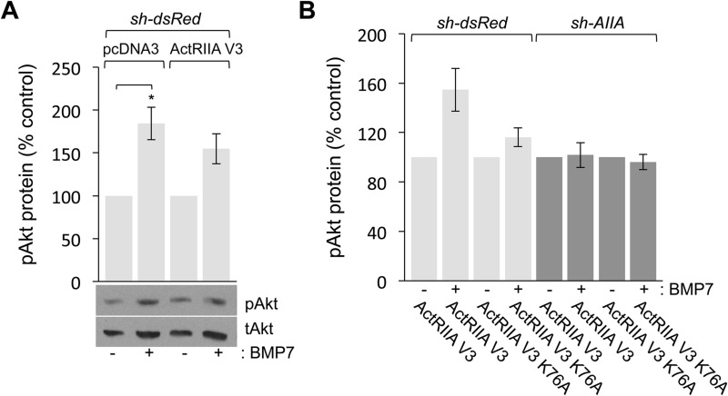
Fig. 7.
BMP7-evoked Akt phosphorylation is inhibited by loss of ActRIIA expression or expression of ActRIIA V3 K76A. (A,B) Quantification (mean±s.e.m.; n=2) of western blots of dsRedΔWEHI and AIIAΔWEHI lysates co-expressing the indicated cDNA expression constructs incubated with or without 50 ng/ml BMP7 for 30 min and probed for pAkt. Detection of tAkt provided a loading control. pAkt levels for each transfection condition were normalized to pAkt levels in the respective unstimulated cells. (A) BMP7 stimulated increases in the levels of pAkt in dsRedΔWEHI cells co-expressing pcDNA3 (84%±19% over control; *P<0.05, Student's two-tailed t-test) or ActRIIA V3 (55%±17% over control). (B) BMP7 stimulated increases in the levels of pAkt only in dsRedΔWEHI co-expressing ActRIIA V3 (55%±17% over control, lane 2). Increases in pAkt over control levels were not observed in any other condition in response to BMP7 including in dsRedΔWEHI co-expressing ActRIIA V3 K76A (16%±7.5% over control, lane 4), AIIAΔWEHI co-expressing ActRIIA V3 (1.8%±4.7% over control, lane 6) or AIIAΔWEHI co-expressing ActRIIA V3 K76A (−3.8%±6.2% over control, lane 8).

