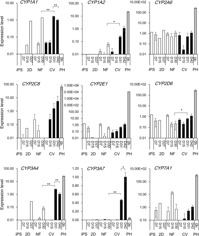
Fig. 3.
Expression of CYP metabolizing enzyme genes in differentiated hiPS cells on the collagen vitrogel or nanofiber matrices or 2D plates. Expression levels of various metabolizing enzymes were quantified by real-time PCR in undifferentiated hiPS cells (iPS) or hiPS-hep, grown on tissue culture plates pre-coated with SynthemaxII (2D), nanofiber (pre-coated with Matrigel; NF) or the CV membranes. Primary hepatocytes (PH, 0 h) were used as a reference. For differentiated hiPS cells, values represent means±s.d. (n=3). Relative values are shown, with the value of D42 hiPS cells grown on the CV=1. *P<0.05 or **P<0.01, by two tailed Student's t-test, compared between hiPS-hep grown on CV (D31) versus NF (D30); or CV (D31) versus CV (D42).

