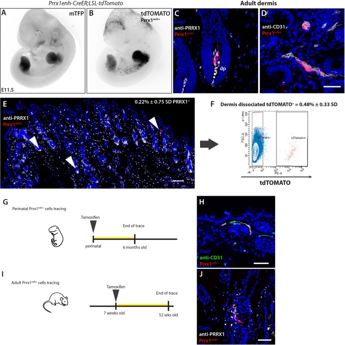
Fig. 2.
The Prrx1 enhancer is active in limb-bud mesenchyme progenitors and adult dermis. To lineage trace the fate of Prrx1enh+ cells, a transgenic Prrx1 enhancer-CreER-T2A-mTFPnls was crossed to a reporter line Rosa-CAG-loxP-stop-loxP-tdTomato (referred to as Prrx1enh-CreER;LSL-tdTomato). (A) Inverted greyscale images of embryos showing limb TFP expression at E11.5 under the Prrx1 enhancer. (B) Upon tamoxifen administration (1 mg oral gavage to mothers 24 h prior to imaging), tdTOMATO+ (Prrx1enh+) is visible in limb buds. Additionally, Prrx1enh+ cells are present in a salt-and-pepper pattern in inter-limb flank, as well as cranial and craniofacial mesenchyme. (C,D) Prrx1enh+ cells in adult limb dermis. Cre recombinase activity labels cells primarily in dermal papilla (dp) and perivascular cells. Scale bar: 50 μm. (E) An overview of the sparsely-labeled cells within the adult limb dermis. Arrowheads mark Prrx1enh+ positive cells that make up 0.22%±0.75 s.d. of PRRX+ cells. Scale bar: 200 μm. (F) Dermal dissociation of cells 3 weeks after tamoxifen administration and FAC sorting of tdTOMATO+ cells shows that in adult skin only a small population of cells retain the activity of the Prrx1 enhancer. (G) Graphic representation of experimental setup to analyze the role of perinatal Prrx1enh+ cells in tissue homeostasis. (H) A representative tissue section of experiment (G) after 6 months. Scale bar: 50 μm. (I) Graphic representation of experimental setup to analyze the role of postnatal Prrx1enh+ in tissue homeostasis. (J) A representative micrograph of experiment (I) after 1 year. Scale bar: 50 μm.

