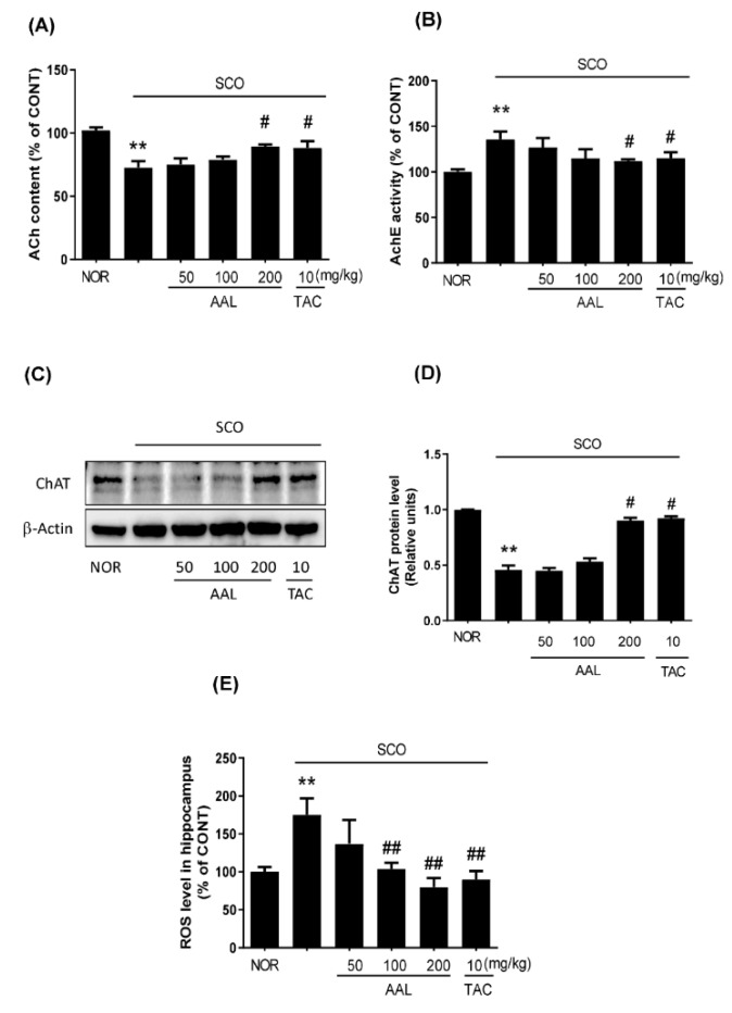
Figure 4.
Effect of AAL extract on cholinergic system dysfunction and ROS production in scopolamine-induced cognitive deficit mouse brains. ACh content (A) and AChE activity (B) were measured in the hippocampus using an ACh and AChE activity assay kit (US Biomax Inc., Rockville, MD, USA). Hippocampus tissue was lysed and subjected to Western blotting with anti-ChAT antibody (C). Expression levels were normalized to β-actin. Bar graphs represent the relative band intensities compared with NOR (D). ROS levels in hippocampal tissue were measured using an ROS/RNS assay kit (Cell Biolabs, Inc., Sandiego, CA, USA). The fluorescence intensities corresponding to the ROS levels in the hippocampus are represented as a percentage of that in the NOR (E). Data are presented as the mean ± SEM (n = 5). ** p < 0.01 vs. NOR group, # p < 0.05 or ## p < 0.01 vs. SCO group. NOR: normal control; SCO: scopolamine; AAL: A. atemoya leaf; TAC: tacrine.

