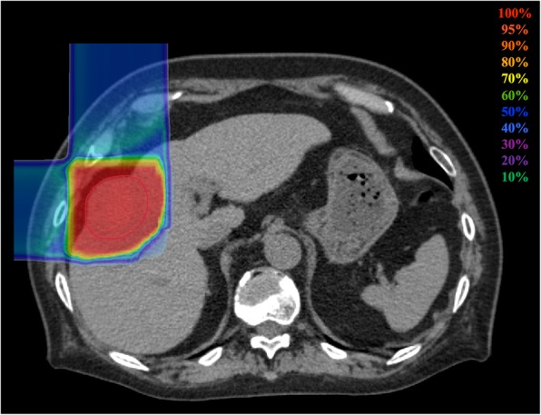
Fig. 1.

Dose distribution of C-ion RT for HCC. Isodose curves of C-ion RT are superimposed on an axial computed tomography image for the total irradiation plan. The area within the red outline is the gross target volume. Highlighted are 100% (red), 95% (light red), 90% (orange), 80% (light orange), 70% (yellow), 60% (green), 50% (blue), 40% (cyan), 30% (light purple), 20% (purple), 10% (light blue) isodose curves (100% was 60 Gy [relative biological effectiveness])
