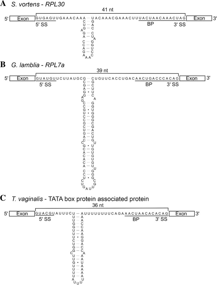
Fig. 3.
Base pairing within long cis-spliced introns in diplomonads and a parabasalid. Secondary structural predictions of representative cis-spliceosomal introns from S. vortens (a), G. lamblia (b), and T. vaginalis (c) are shown with putative 5′/3′ splice site (SS) and branch point (BP) motifs underlined. Lengths of ‘single-stranded’ distances between splice donor and acceptor sites are indicated in nucleotides (nt) above the intron sequences

