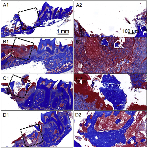
Fig. 6.
Masson’s trichrome-stained images of different experimental groups studied for periodontal bone healing. (A1/A2) Immediately after tooth extraction and defect creation, (B1/B2) Unfilled defect/control after 4 weeks of surgery, (C1/C2) Mineralized PCG nanofiber fragments after 4 weeks of graft implantation, and (D1/D2) E7-BMP-2- loaded mineralized PCG nanofiber fragments after 4 weeks of graft implantation. The dashed boxes indicate the defect region.

