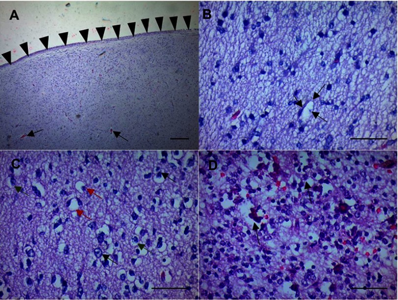
Figure 2.
In postmortem microscopic analysis of samples obtained from the fetus autopsy. (A) The sulci have almost disappeared, making the brain surface smooth, which is highlighted in the image by the small triangles and the arrows showing intensive gliosis with Rosenthal fibers. (B) Neural loosening due to the neural injury in the surroundings. It is possible to see some inflammatory cells and neural vacuolation (arrows). (C) Arrow shows non-specific reactive changes with astrocyte proliferation, gliosis, and edema (astrogliosis) as part of the glial scaring process. Neural vacuolation and degenerate axons devoid of myelin sheath (Wallerian degeneration, red arrows) and macrophages occupying the space of a former axon (green arrows). (D) Microcalcifications (arrows) and gliosis. Hematoxylin and eosin staining was used to visualize the previous panels. Scale bar is shown on the bottom right of each panel. Scale bar is shown on the bottom right of each panel. Scale bar for figure A 1.03 um/pixel and figures B, C and D 0.518 um/pixel.

