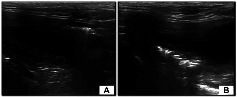
Figure 3.
Longitudinal ultrasonographic scan of the urinary bladder of a 4-year-old female English setter dog in dorsal recumbency (A) and in standing position (B). Note the hyperechoic interface with reverberation artifacts moving to the dorsal wall according to the change of position. The dog had chronic kidney disease complicated with UTI (E. coli was cultured from the urine), with no evidence of diabetes mellitus.

