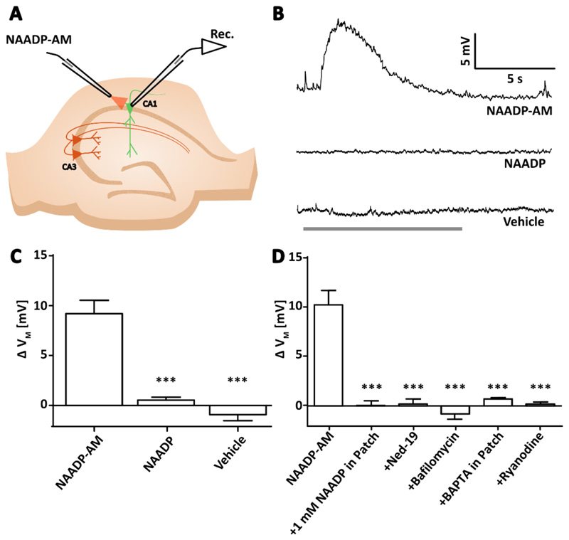
Figure 1. NAADP causes membrane depolarization in pyramidal neurons of the hippocampus in a manner dependent on acidic store signaling, intracellular Ca2+, and RyR.
(A) Diagram showing the experimental configuration to record membrane potential of CA1 or CA3 pyramidal neurons in hippocampal slices whilst NAADP-AM was applied locally. (B) Example voltage traces recorded whilst applying NAADP-AM, NAADP, or vehicle. Arrows indicate the start of delivery and grey bar indicates the total time of application. (C) Transient membrane depolarization (ΔVM) upon application of NAADP-AM (300 μM, n=12 cells), NAADP (300 μM, n=5), or vehicle (n=6). Data are mean ±SEM. (D) Mean ΔVM upon application of NAADP-AM (300 μM, n=11 cells) alone or (left to right) in combination with a desensitizing concentration of NAADP (1 mM) inside the internal solution of the patch pipette (n=6); preincubation with the NAADP antagonist Ned-19 (100 μM, 40 min, n=6); preincubation with the vacuolar H+ ATPase inhibitor bafilomycin (4 μM, 40 min, n=5); with BAPTA (15 mM) inside the internal solution of the patch pipette (n=5); or pre-incubation with ryanodine (30 μM, 40 min, n=4). Significance was assessed with; Data are mean ±SEM; n = single cells. *** P < 0.005 by Kruskal-Wallis and post hoc Dunn’s tests.

