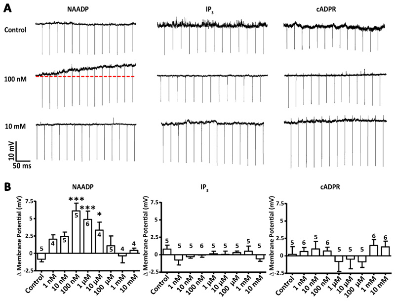
Figure 2. NAADP is unique among second messengers in its ability to depolarize hippocampal pyramidal neurons.
(A) Example voltage traces for dialysis of CA1 pyramidal neurons patched with internal solutions containing various concentrations of the Ca2+- mobilizing second messengers NAADP, IP3 and cADPR. Changes to membrane potential were recorded over time as the second messengers dialysed into the patched cell. (B) Transient membrane depolarization (ΔVM) of the cells described in (A) in response to increasing concentrations of the Ca2+-mobilizing second messengers. Data are mean ± SEM; n = single cells, indicated above each column; n > 4 for all concentrations of second messengers. *** P < 0.005 and * P <0.05 by Kruskal-Wallis and post hoc Dunn’s tests.

