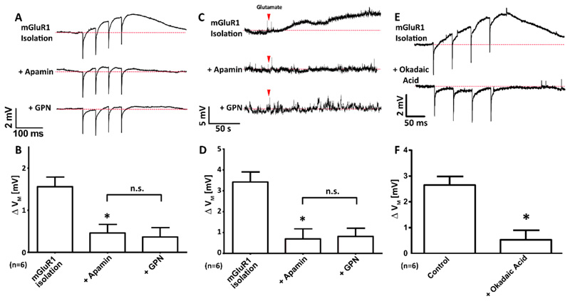
Figure 5. In CA1 pyramidal neurons, mGluR1-dependent depolarization occurs through the inactivation of SK channels by possibly protein phosphatase 2A (PP2A).
(A and B) Representative voltage recordings (A) and mean Δ VM (B) upon electrical stimulation (4x, 20 Hz) of CA1 neurons whilst mGluR1 was pharmacologically isolated, then subsequent addition of apamin (200 nM, 15 min) and finally GPN (200 μM, 10 min); n=6 cells. (C and D) Representative voltage recordings (C) and mean Δ VM (D) upon bath application of glutamate (red arrowhead; 300 μM, 120 s) of CA1 neurons whilst mGluR1 was pharmacologically isolated, then subsequent addition of apamin (200 nM, 15 min) and finally GPN (200 μM, 10 min); n=6 cells. (E and F) Representative voltage recordings (E) and mean Δ VM (F) upon electrical stimulation (4x, 20 Hz) of CA1 neurons whilst mGluR1 was pharmacologically isolated in the absence or presence of okadaic acid (100 nM, 15 min); n=6 cells. Data are means + SEM, each from n = 6 single cells. ** P < 0.01 and * P < 0.05 (relative to mGluR1 isolation-alone condition), and n.s. = no significant difference; by Freidman’s test with post hoc Dunn’s tests (B and D) or a Wilcoxson pair matched signed ranks test (F).

