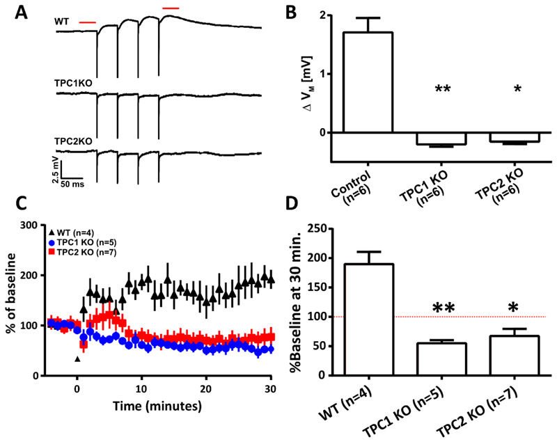
Figure 7. In CA1 pyramidal neurons, Two-pore channels are required for mGluR1-mediated membrane depolarization and mGluR1-dependent LTP.
(A) Representative average (5 traces) voltage recordings from CA1 neurons in hippocampal slice preparations from wild-type (WT), Tpc1-/-, and Tpc2-/- mice (n=6 mice each). Recordings were obtained upon pharmacological isolation of mGluR1 (50 μM AP5, 10 μM NBQX, 100 nM LY341495, 10 μM MPEP, 100 μM picrotoxin, and 2 μM CGP 55845) and electrical stimulation (4x, 20 Hz) of afferent fibres in stratum radiatum. Dashed red line indicates where membrane potentials were compared before and after stimulation. (B) Columns show mean ΔVM of CA1 pyramidal neurons before and after electrical stimulation described in (A). (C) A causal spike-time-dependent plasticity protocol was used to induce mGluR1-dependent LTP after a baseline of EPSPs were recorded for 5 min (indicated by marker at 0 min) in WT (n=4), Tpc1-/- (n=5) and Tpc2-/- (n=7) animals. One casual presynaptic stimulation is paired with two backpropagating action potentials (100 Hz) at a 10 ms interval. (D) Mean change in synaptic strength at 25-30 min shown/described in (C), expressed as a % of the baseline (red dashed line). Data are means ± SEM, n = single cell. ** P < 0.01 and * P < 0.05 by Kruskal-Wallis and post hoc Dunn’s tests.

