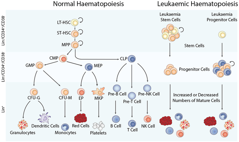
Figure 1. Comparison of normal versus leukaemic haematopoiesis.

During normal haematopoiesis (left), a small number of haematopoietic stem cells gives rise to a limited number of progenitor cells, which subsequently gives rise to mature members of all blood cell types, including red blood cells, myeloid cell lineages such as granulocytes, dendritic cells, monocytes, or platelets, and lymphoid lineage cells, including NK cells, and T and B lymphocytes. In malignant haematopoiesis (right), leukaemic stem and progenitor cells with enhanced self-renewal or proliferation and reduced apoptosis give rise to abnormal numbers of mature cells in the bone marrow, peripheral blood, and other tissues. CFU-G, colony forming unit-granulocyte; CFU-M, colony forming unit-monocyte; CMP, common myeloid progenitor; EP, erythroid progenitor; GMP, granulocyte-macrophage progenitor; LT-HSC, long term-haematopoietic stem cell; MEP, megakaryocyte-erythroid progenitor; MKP, megakaryocyte progenitor; MPP, multipotent progenitor; NK cell, natural killer cell; ST-HSC, short term-haematopoietic stem cell.
