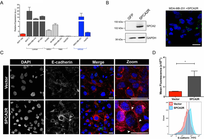
Figure 3. SPCA2 increases E-cadherin expression in TNBC cells.
(A) A panel of breast cancer cell lines classified as luminal, HER2+ or TNBC subtypes as indicated, was evaluated for SPCA2 expression by qPCR. Results are normalized to MCF-10A. SPCA2 transcripts were lowest in two TNBC lines, MDA-MB-231 and Hs578T, and could be significantly increased by lentiviral-mediated transfection of SPCA2R. (B) SPCA2R expression in transfected MDA-MB-231 cells was confirmed by Western blotting (left) and confocal immunofluorescence microscopy (right) using anti-FLAG antibody. (C) MDA-MB-231 Tet-Ecadherin cells expressing either vector or SPCA2R were treated with 2μg/ml of doxycycline, stained for E-cadherin and imaged using confocal microscopy. (D) Normalized fluorescence values for each condition (top) and histograms of cells separated by flow cytometry (bottom). *p<0.05, Student’s t-test, vector n=3, SPCA2R n=3. Flow cytometry histogram analysis of 3 biological replicates of each condition. Vector n=9468 cells, SPCA2R n=9258 cells. See Fig. S3.

