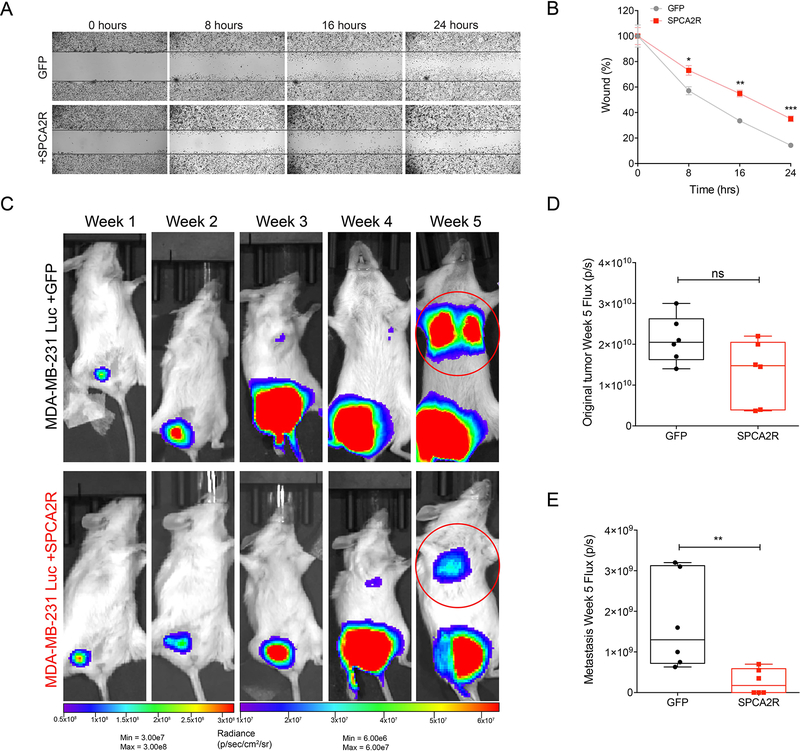
Figure 7. SPCA2 suppresses migration in vitro and metastasis in vivo.
(A) Representative images of wound healing in MDA-MB-231 at the indicated times in control and SPCA2R transfected cells (2.5x magnification). (B) Quantification of cell migration expressed as percentage of control. SPCA2 significantly reduced migration at 8 h, 16 h, and 24 h. *p<0.05, **p<0.01, ***p<0.01. Student’s t-test, n=3 for each condition. (C) Bioluminescent images of NSG mice following engraftment with MDA-MB-231 Luc + GFP and MDA-MB-231 Luc + SPCA2R at indicated times and luminescence scales. (D-E) Quantification of bioluminescence in luciferase-labeled MDA-MB-231 Luc. Tumor growth and metastasis at Week 5 were represented as mean total flux (p/sec) ± SEM. SPCA2 significantly reduced metastasis compared with control group. **p<0.01, Student’s t-test, n=6 for each condition. See Fig. S7.

