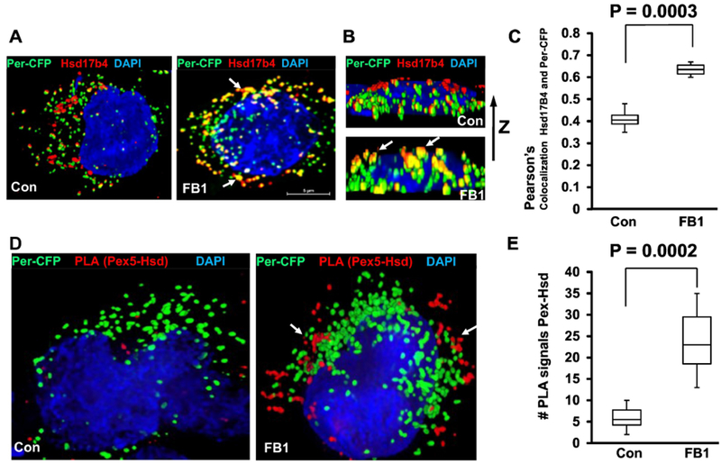
Figure 5. Ceramide depletion induces translocation of Hsd17b4 to peroxisomes and interaction with Pex5.

A-C. HEK293T cells expressing the peroxisome marker Per-CFP (green) were depleted of ceramide by FB1 treatment and subjected to immunocytochemistry using antibody against Hsd17b4 (red). A, top view; B, side view. Arrows point at peroxisomes co-localized with Hsd17b4. C, Quantitative analysis (Pearson’s coefficient) for colocalization of Per-CFP with Hsd17b4. N=5, P<0.01. D, E. PLAs using antibodies against Pex5 (rabbit IgG) and Hsd17b4 (mouse IgG) with HEK293T cells expressing Per-CFP (green). E is quantification of D for number of PLA signals. N=4.
