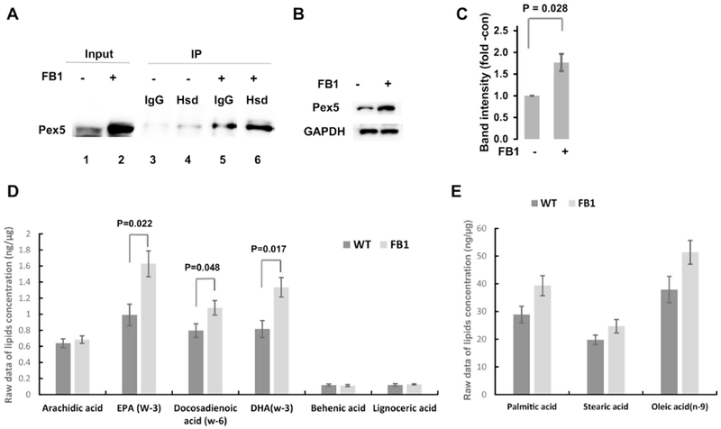
Figure 6. Ceramide depletion by FB1 treatment upregulates Pex5 and generation DHA and EPA.

A. Coimmunoprecipitation analysis using cell lysates of HEK293T cells with or without prior treatment with 5 μM FB1 for 48 h. Antibody against Hsd17b4 was used for immunoprecipitation (IP) and anti-Pex5 antibody was used for the immunoblotting reaction (IB). Lanes 1, 2: input; lanes 3, 4: output without FB1; lanes 5, 6: output with FB1. FB1 treatment increases level of Pex 5 in input and output. IgG, non-specific mouse IgGused or IP. B, C. SDS-PAGE/immunoblot for protein levels of Pex5 in different cell types/tissue with or without FB1 treatment as in A. C is quantitation of B. N=5. P<0.05 as indicated in figure. D, E. Mass spectrometric analysis of free fatty acids in HEK293T cells with or without FB1 treatment as in A. D, 3-PUFAs and VLCFAs. E. Medium chain fatty acids. N=5. P<0.05 as indicated in figure.
