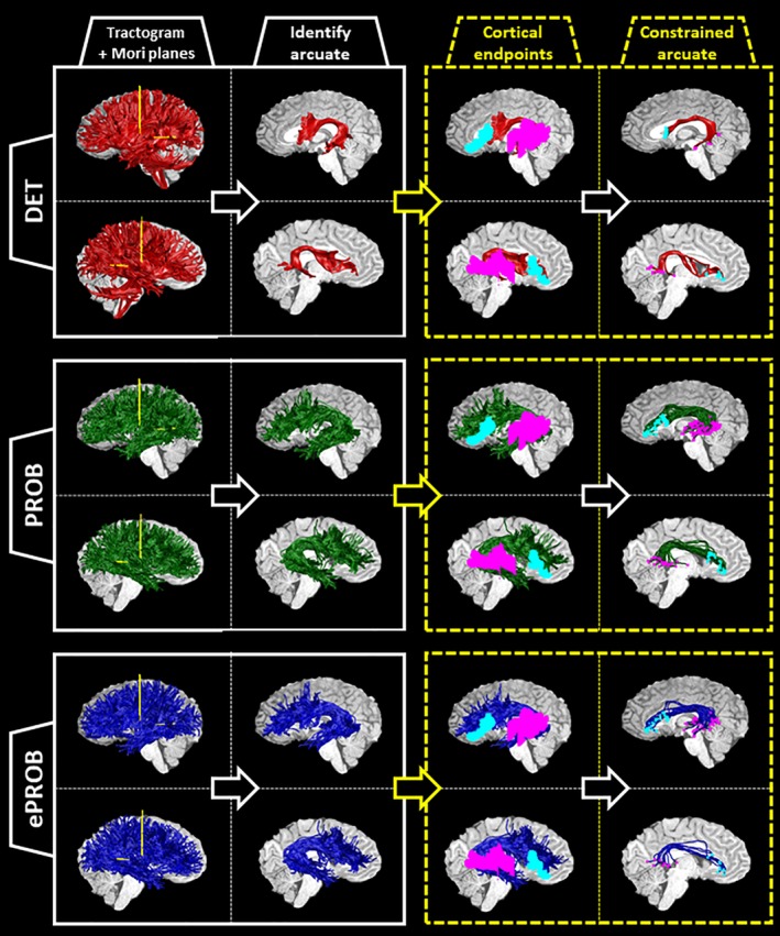
Figure 1.

Pipeline for identifying the arcuate fasciculus from three whole‐brain tractograms, illustrated on a sample subject. For each subject, a whole‐brain set of streamlines is generated three times: (1) with deterministic, tensor‐based tracking, labeled DET (red); (2) with a probabilistic, spherical‐deconvolution–based tracking, labeled PROB (green); and (3) with the application of a global evaluation algorithm to reduce the PROB tractogram, labeled ePROB (blue). After the tractogram is generated, two Mori waypoint planes (yellow) are inserted into each hemisphere (first column; see also Figure A11). The streamlines that intersect both planes in each hemisphere are labeled arcuate streamlines (second column). To incorporate additional anatomical information about the cortical destinations of the arcuate, we choose only those arcuate streamlines that connect to both the frontal and temporal cortical regions of interest (ROIs; cyan and magenta, respectively, in the third column). By implementing this constraint, we define a new, subset arcuate bundle (last column, with the endpoints marked in cyan and magenta). For each tractography, we show the left hemisphere on top and the right hemisphere on the bottom [Color figure can be viewed at http://wileyonlinelibrary.com]
