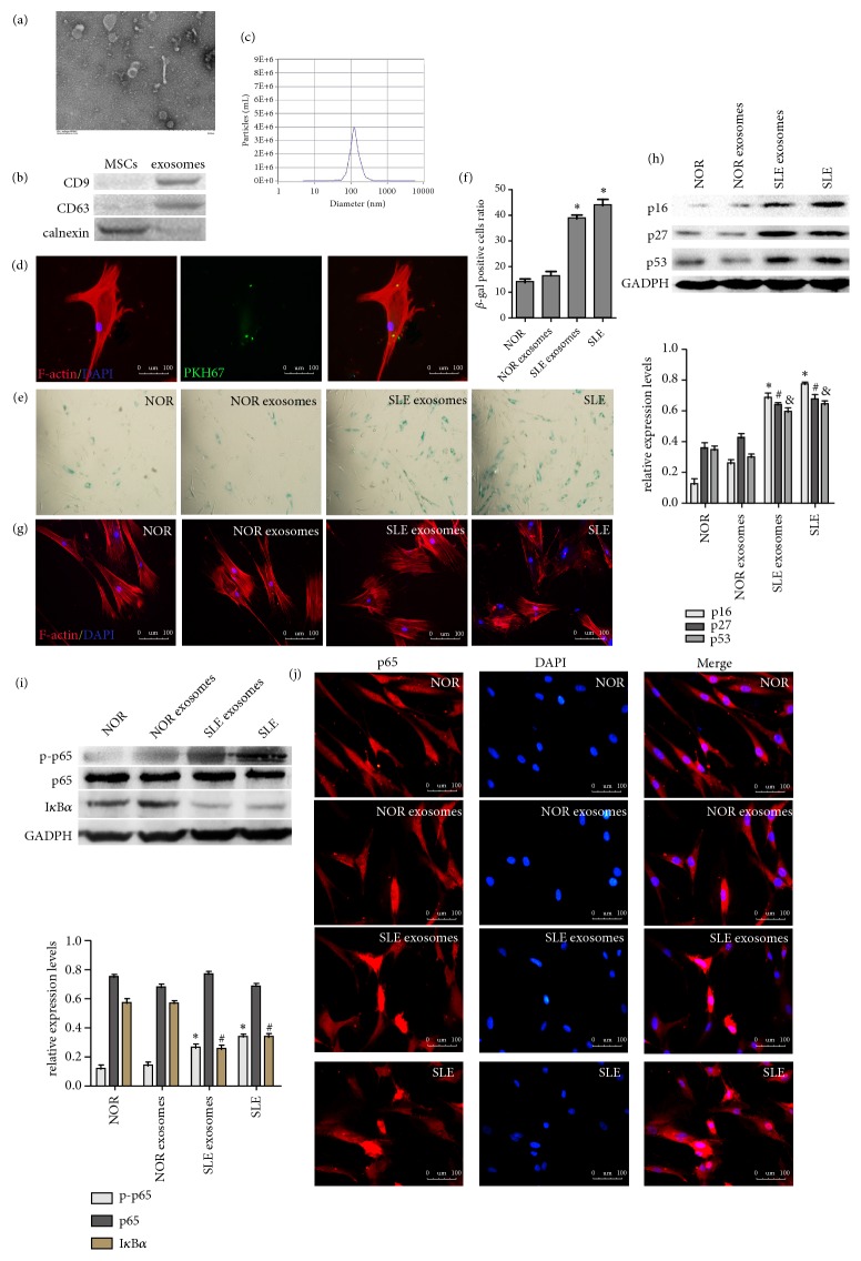
Figure 2.
Serum exosomes from SLE patients enhanced the senescence of MSCs by activating NF-κB signaling pathway. (a) Serum exosomes were observed under a transmission electron microscope. (b) Western blotting analysis of CD9, CD63, and calnexin expression in lysates from purified serum exosomes. (c) Nanoparticle tracking analysis was used to detect the size distribution of exosomes. (d) BM-MSCs were incubated in serum exosomes that were labeled with PKH67 (green). (e, f) The number of SA-β-gal-positive cells was increased in the SLE serum exosomes-treated BM-MSCs in comparison with the normal group. (g) Immunofluorescence showed that distribution of F-actin in the BM-MSCs was disordered after being stimulated with SLE exosomes. (h) The protein expressions of p16, p27, and p53 were increased in MSCs treated with SLE serum exosomes by western blotting analysis. (i) Western blot analysis was performed to examine the protein expressions of p-p65, p65, IκBα. (j) Immunofluorescence staining of p65 in BM-MSCs treated exosomes (∗P < 0.05 compared with the normal group, #P < 0.05 compared with the normal group, &P < 0.05 compared with the normal group) (NOR: normal MSCs group, NOR exosomes: normal MSCs treated with normal human exosomes, SLE exosomes: normal MSCs treated with exosomes from SLE patients, SLE: SLE MSCs group).

