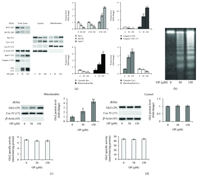
Figure 1.
The proapoptotic effect of OP is associated with increased expression of mitochondrial Glo2 in NSCLC A549 cells. (a) Antiapoptotic Bcl-2 and Bcl-XL, proapoptotic Bax, cytochrome c (Cyt c), Apaf-1, and caspase-3 (intact protein, 32 kDa molecular weight; active fragments, 20 and 17 kDa molecular weight) protein expression in untreated (0 μM) and oleuropein- (OP-) treated (50 and 150 μM) A549 cells. (b) Apoptosis was confirmed at morphological level by DNA fragmentation, evaluated by gel electrophoresis. Electrophoresis is a representative of three independent experiments providing the same result. Evaluation of Glo2 expression, by western blot, and enzyme specific activity, by a spectrophotometric assay, in the (c) mitochondria and (d) cytosolic lysates of A549 cells. Histograms indicate the means ± SD of three different cultures, each of which was tested in quadruplicate and expressed as a percentage of control. Western blot analysis of β-actin or CoxIV expression is provided to show equal loading of the samples and to demonstrate successful enrichment of mitochondria in fractionated extracts. Blots are representative of three independent experiments, which gave the same results. ∗p < 0.05, ∗∗p < 0.01, and ∗∗∗p < 0.001, significantly different from control untreated cells.

