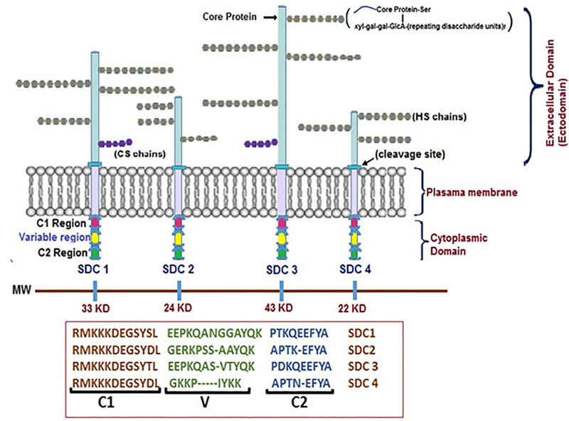
FIGURE 1.
Structural organization of Syndecans. A schematic view of syndecan core protein and glycosaminoglycan chains. SDC-1 and SDC-3 core proteins are larger than SDC-2 and SDC-4 and in addition to heparan sulfate chains they also bear CS. The GAG chains are substituted on core protein serine residues and have common tetrasaccharide units attached to two units of galactose (gal) and GlcA residue with alternating units of GlcNAc and uronic acids. The HS chains undergo modification by sulfonation at 6-O or 3-O (rarely), CS 6-O, or 4-O while uronic acids undergo epimerization to IdoA. The cytoplasmic domain contains highly conserved regions (C1 and C2) with interceding variable (V) regions

