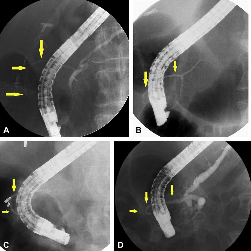
Figure 5.

A.) Ventral pancreatogram demonstrating annular duct (yellow arrows), with otherwise normal ductal anatomy (i.e. no pancreas divisum). B and C.) Ventral pancreatogram demonstrating annular duct (yellow arrows), with pancreatic divisum anatomy (no communication with main pancreatic duct). D.) Ventral pancreatogram demonstrating incomplete annular duct (yellow arrows) and findings of main duct suspicious for chronic pancreatitis including dilated main duct and prominent and irregular side branches.
