Figure 2.
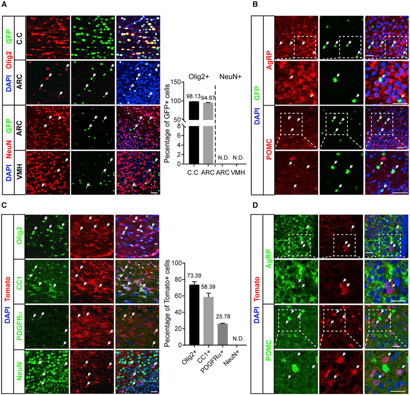
Gpr17 Is Expressed Predominantly in the Oligodendrocyte Lineage (A) Immunofluorescent analysis of GFP, Olig2, and NeuN in cryostat sections of the corpus callosum (C.C), arcuate nucleus (ARC), and ventromedial nucleus (VMH) of Gpr17−/− mice as described in STAR Methods. Scale bar, 25 μm. The percentage of Olig2- or NeuN-positive cells was quantified as indicated. N.D., not detected.
(B) Immunofluorescence analysis of GFP and AgRP or POMC in the ARC of Gpr17−/− mice. Scale bar, 25 μm. Arrows indicate GFP-positive cells.
(C) Immunofluorescence analysis of tdTomato and oligodendrocyte lineage markers, including Olig2, CC1, and PDGFRα, and the neuron marker NeuN in cryostat sections from the C.C (Olig2, CC1, and PDGFRα) or ARC (NeuN) in Olig1cre/+;tdTomato mice. Scale bar, 25 μm. Arrows indicate tdTomato-positive cells. The percentages of Olig2-, CC1-, PDGFRα-, and NeuN-positive cells were quantified as indicated.
(D) Immunofluorescence analysis of tdTomato and AgRP or POMC in the ARC of Olig1cre/+;tdTomato mice. Scale bar, 25 mm. Arrows indicate tdTomato-positive cells.

