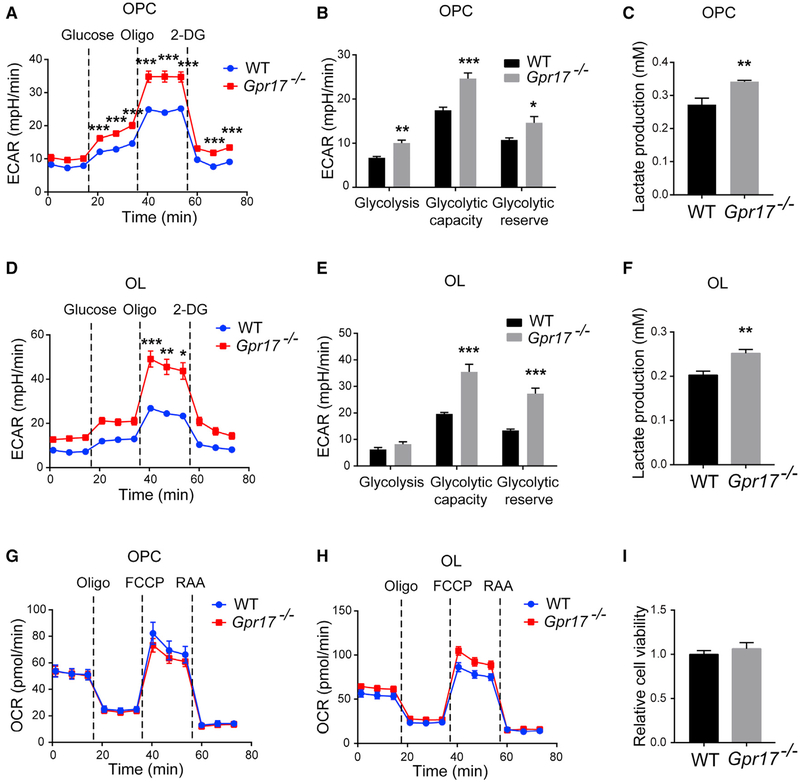
Figure 4.
Ablation of Gpr17 Promotes Glycolysis in Oligodendrocytes Primary OPCs from WT or Gpr17−/− mice were isolated, cultured, and differentiated into oligodendrocytes (OLs) as described in the STAR Methods.
(A) ECAR of WT and Gpr17−/− OPCs as a function of time after sequential administrations of 10 mM glucose, 1.5 μM oligomycin, and 50 mM 2-Deoxy-D-glucose (2-DG). Dotted lines indicate the starting point of treatment with the indicated compounds.
(B) Quantifications of the ECAR results in (A).
(C) Lactate concentration in the medium of WT and Gpr17−/− OPCs.
(D) ECAR of WT and Gpr17−/− OLs.
(E) Quantifications of the ECAR results in (D).
(F) Lactate concentrations in the culture medium of WT and Gpr17−/− OLs.
(G and H) OCR was monitored over time after sequential administration of 1.5 μM oligomycin, 1.5 μM carbonyl cyanide-4-(trifluoromethoxy)phenylhydrazone (FCCP), and 0.5 mM rotenone and antimycin A mixture in (G) OPCs and (H) OLs.
(I) Viability of mouse OPCs measured with the CCK-8 assay.
Data are means of at least three independent experiments. Each value represents mean ± SEM. *p < 0.05, **p < 0.01, ***p < 0.001, Student’s t test.

