
Figure 1. Treg-Specific Deletion of Tfam Results in Severe Systemic Inflammation in Mice.
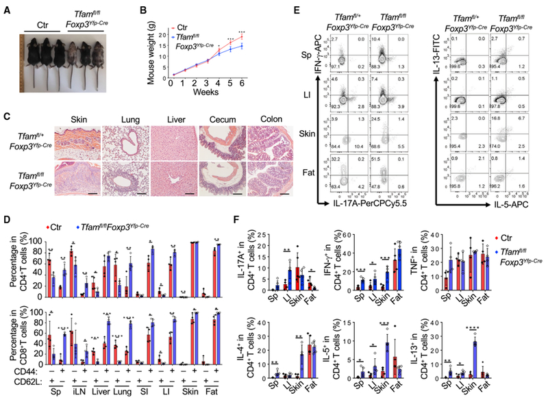
Genotypes of the control (Ctr) group include Tfam+/+Foxp3Yfp-Cre and Tfamfl/+Foxp3Yfp-Cre unless otherwise noted.
(A) Representative figure of 6-week-old mice of the indicated genotypes.
(B) Tfam conditional knockout by Foxp3Yfp-Cre resulted in lower body weight from 4 weeks of age. Ctr, n = 6; Tfamfl/flFoxp3Yfp-Cre, n = 5.
(C) H&E staining of multiple organ and tissue sections from 6-week-old mice of the indicated genotypes. Data are representative of Ctr (n = 3) and Tfamfl/flFoxp3Yfp-Cre (n = 2) mice.
(D) Percentages of CD44 and CD62L expression in CD4+ (top) and CD8+ (bottom) T cells in different organs of 6-week-old Ctr (n = 4) and Tfamfl/flFoxp3Yfp-Cre (n = 4) mice.
(E) Flow cytometry analysis of IL-17A, IFN-γ, IL-5, and IL-13 expression in CD4+ T cells in the spleen (Sp), large intestine (LI), skin, and fat of 6-week-old mice of the indicated genotypes. Data are representative of three independent experiments.
(F) Percentages of IL-17A, IFN-γ, TNF, IL-4, IL-5, and IL-13 expression in CD4+ T cells in 6-week-old Ctr (n = 5) and Tfamfl/flFoxp3Yfp-Cre (n = 5) mice. Data are shown as mean ± SD in (B), (D), and (F).