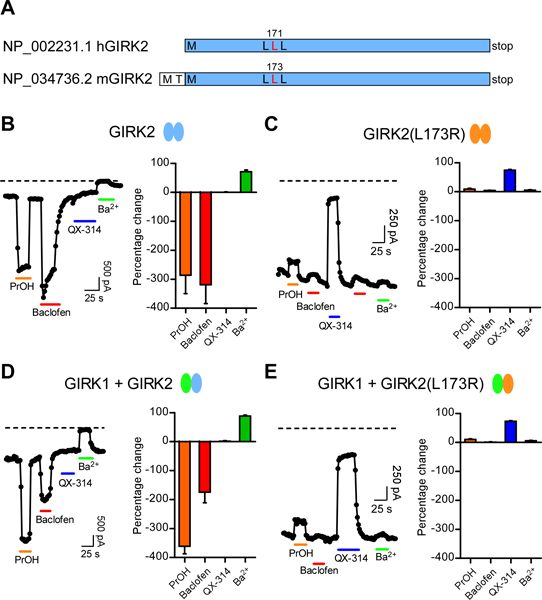
Figure 3: Change in gating and ion selectivity of mouse GIRK2(L173R) channels.

(A) Cartoon shows similarity between human and mouse GIRK2 protein, with homologous Leu highlighted (red). (B,C) Whole-cell patch-clamp currents are plotted as a function of time for wild-type GIRK2 (B) and mutant GIRK2(L173R) (C) channels. Currents were measured at −120 mV from HEK293T cells transfected with GABAB1b/B2 receptor cDNA and either GIRK1, GIRK2 and/or GIRK2(L173R) cDNAs. Plots show the response to bath application of PrOH (100 mM, orange bar), Baclofen (100 μM, red bar), the sodium channel inhibitor QX-314 (100 μM, blue bar) and the Kir potassium channel inhibitor Ba2+ (1 mM, green bar). Note that the mutant GIRK2(L173R) channel shows no activation with Baclofen or PrOH, lack sensitivity to inhibition with Ba2+, but is inhibited with QX-314 (100 μM). Similar results were observed for HEK293T cells expressing heterotetramers of GIRK1+GIRK2 (D) or GIRK1+GIRK2(L173R). (E) channels. Dashed line indicates zero current level. Bar graphs show average percentage change (± SEM) in current with indicated compounds (N=8–9). A negative percentage change indicates activation.
