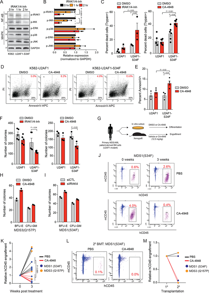
Figure 6. U2AF1-S34F AML cells are sensitive to IRAK4 inhibitors.
(A) K562-U2AF1-S34F cells were treated with 10 mM IRAK1/4-inhibitor for 1 or 2 hours and immunoblotted for NF-κB and MAPK activation. (B) Densitometric analysis of panel (A) summarized from three independent biological replicates. Two-sided t-tests were used for statistical analyses. (C) K562-U2AF1 cells were treated with DMSO, 10 μM IRAK1/4-Inh, or 10 μM CA-4948 for 7 days and assessed for viability by Trypan Blue exclusivity (three independent experiments). One-sided t-tests were used for statistical analyses. (D) Representative images of K562 cells expressing wild-type U2AF1 or U2AF1-S34F were treated with DMSO or 10 μM CA-4948 for 48 hours days and then analyzed by flow cytometry for AnnexinV and Propidium Iodide (PI) staining (three independent experiments). (E) Summary of (D). Two-sided t-tests were used for statistical analyses.(F) K562 cells treated with 10 mM IRAK1/4-Inh (five independent experiments) or CA-4948 (three independent experiments) were evaluated after 7 days for colony formation in methylcellulose. Two-sided t-tests were used for statistical analyses. (G) Schematic of experimental design. (H) MDS patient-derived BM cells were evaluated after 7 days for colony formation in methylcellulose treated with 0.5 μM CA-4948 or vehicle (two independent experiments). (I) MDS patient-derived BM cells were transfected with siIRAK4 or control siRNA and then evaluated after 7 days for colony formation in methylcellulose (two independent experiments). (J) NSG mice were xenografted with U2AF1-mutant MDS BM cells. After engraftment, mice were treated with CA-4948 or vehicle and human cell engraftment was assessed by flow cytometry on BM aspirates. (K) Summary of BM cell engraftment (hCD45) for independent mice and patient-derived samples. Two-sided t-tests were used for statistical analyses. (L) MDS BM cells were isolated from mice treated with vehicle (PBS) or CA-4948 treatment after 3 weeks (panel J,K) and then xenografted into secondary NSG mice. Human cell engraftment was assessed by flow cytometry on BM aspirates. (M) Summary of BM cell engraftment (hCD45) for mice and 2 independent patient-derived samples. Data represent the mean ± s.e.m. for all panels except H and I where data represent the mean ± s.d.

