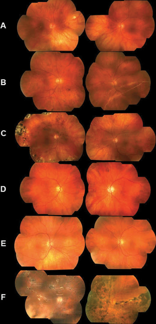
Figure 2.
Fundus photos of affected individuals harboring the p.Gln39* mutation. (A) Photos of a 42-year-old female (III:3). The right eye underwent retinal detachment repair with scleral buckle at age 24. The vitreous was optically empty in both eyes. In the right eye, there were focal chorioretinal scars in the macula and mid-peripherally in the 2:00 meridian. Not visualized were inferior and temporal vitreous condensation and vitreous sheets bilaterally, as well as indentation from a scleral buckle in the right eye and chorioretinal scarring bilaterally. (B) Photos of the right eye of a male individual III:17 at age 43 (left image) and at age 46 (right image). At age 43, the right eye had an optically empty vitreous centrally with avascular sheets in the far temporal periphery. At age 46, the patient presented after one week of sudden visual field loss. A large retinal detachment could be observed, extending from the 12:00 to the 8:00 meridian, caused by a peripheral retinal hole at 02:00. (C) Photos of a 45-year-old female (III:21) showed peripheral scarring in the right eye from repair of a superior giant retinal tear occurring at the age of 15. The tear was treated with laser and a scleral buckle. Later, an inferior retinal hole was diagnosed and treated with laser and cryotherapy. Both eyes had an optically empty vitreous. (D) Photos of a 69-year-old male (II:2). The fundus exam was normal for the right eye. The left eye had an optically empty vitreous with avascular sheets in the periphery. Outside the extent of the left image was superior retinal scarring from the treatment of two retinal tears with pneumatic retinopexy. (E) Photos of a 66-year-old male (II:6) showed an optically empty vitreous in both eyes with avascular sheets in the far periphery. There was no central chorioretinal scarring, but the left eye had some scarring in the far superior periphery (not shown). (F) Photos of a 58-year-old male (II:11). The right eye sustained several retinal tears and detachments and was treated on multiple occasions with laser, cryotherapy, scleral buckle, vitrectomy, and finally placement of silicone oil. Multiple retinal scars were observed as well as light reflections from the oil. The left eye exhibited extensive chorioretinal scarring and pigmentation after treatment of the retinal detachment and scleral buckle placement.

