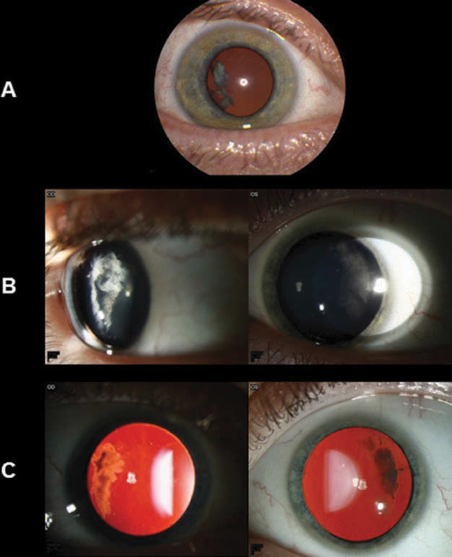
Figure 3.
Slit-lamp and retro-illumination photos of a father (III:18) and son (IV:17) with lamellar cataract in the temporal cortex of each eye. (A) Slit-lamp photo of the 42-year-old father’s right eye, which shows a temporal cortical cataract. The left eye underwent cataract extraction and intraocular lens placement for a visually significant cataract a year previously. (B) Slit-lamp photos of the 12-year-old son’s eyes, which show temporal cortical cataracts that are not visually significant. (C) Retro-illumination of the son’s eyes shows the distinctive temporal location of the cataracts.

