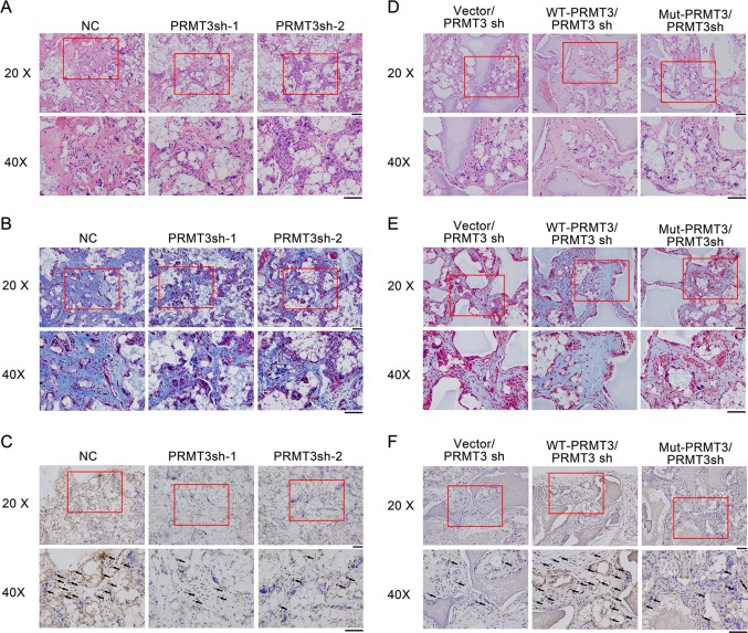
Fig. 3. PRMT3 is a positive regulator of MSC-mediated bone formation in vivo.
a H&E staining of PRMT3 knockdown cells. b Masson’s trichrome staining of PRMT3 knockdown cells. c IHC staining for OCN in PRMT3 knockdown cells. d H&E staining of PRMT3 rescue cells. e Masson trichrome staining of PRMT3 rescue cells. f IHC staining for OCN in PRMT3 rescue cells. H&E staining, hematoxylin-eosin staining; IHC staining, immunohistochemistry staining; Scale bars represents 50 μm; Black arrows indicate positive staining of OCN; n = 6

