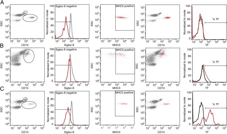
FIGURE 1.
Flow cytometric gating strategy for separating monocytes from granulocytes and eosinophils. (A) Nonstimulated blood. Monocytes were easily distinguished as the SSC+(medium)/CD14+(high), Siglec-8−, and MHCII+ population (red color in fourth picture). (B) CC-stimulated blood. Monocyte and eosinophil populations merged in the CD14+/SSC+ plot; thus, a large gate was set to include the most granular monocytes. Thereafter, monocytes were selected by a negative selection of the Siglec-8− population (based on settings from nonstimulated blood) and a positive selection of the MHCII+ cells (red color, second and third pictures), resulting in a distinguished monocyte population (red color in fourth picture). (C) E. coli–stimulated blood. Monocytes and granulocytes were separated, and the monocytes were distinguished further by Siglec-8− and MHCII+ staining. The percentage of TF+ cells was determined by a histogram plot of fluorescence intensity, determining baseline setting from nonstimulated blood, and the isotype control was subtracted from the positive staining obtained from the specific TF Ab. The granulocytes and eosinophils were further distinguished by negative and positive selection, respectively, of Siglec-8–stained cells (data not shown) and further analyzed for TF expression.

