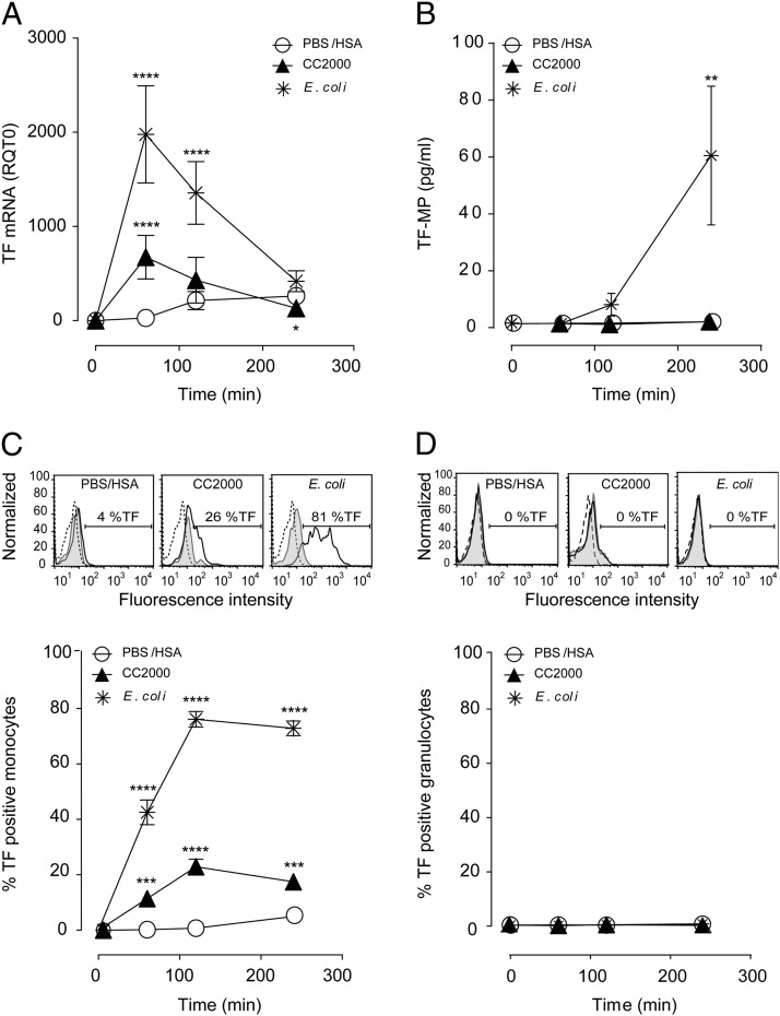
FIGURE 3.
CC induce TF mRNA and TF expression on the monocyte surface. Human whole blood was incubated with CC (500, 1000, and 2000 μg/ml) or controls, PBS/has, or E. coli (1 × 10⁷ particles/ml) at different time points (60, 120, and 240 min). (A) TF mRNA in whole blood measured by quantitative RT-PCR (n = 6 donors); additionally, CC 1000 and 500 μg/ml are in Supplemental Table II. (B) The procoagulant activity of TF-MP (n = 3 donors). (C) Monocyte TF expression exemplified with flow cytometry histogram after 120 min and quantified as percentage of monocytes expressing surface TF at all time points (n = 6 donors). Additionally, CC 1000 and 500 μg/ml are shown in Supplemental Table II. (D) The lack of granulocyte TF expression exemplified with flow cytometry histogram after 120 min and quantified as percentage of granulocytes expressing surface TF at all time points (n = 6 donors). Histogram baselines (T0) are stippled lines, isotype controls are filled gray, and the TF expression are black lines. Graphs are given as means ± SEM. Significant differences between the stimuli and inhibitors at the given time points are marked with asterisks. **p < 0.01, ***p < 0.001, ****p < 0.0001.

